Technical Support for Cloning Products
General information
Formation of disulfide bonds
Some vectors provide a N-terminal fusion of the ompA signal peptide which mediates the secretion of the recombinant protein to the periplasmic space of E. coli. There, the signal peptide is selectively cleaved by the E. coli signal peptidase. The secretion strategy is essential for the functional production of proteins containing structural disulfide bonds that are often present in naturally secreted proteins. The reducing conditions in the cytoplasm of E. coli prevent disulfide bond formation which can lead to aggregation or degradation of unfolded polypeptides.
Periplasmic secretion as a first purification step
Furthermore, periplasmic secretion separates the recombinant protein from cytosolic proteases. Since the E. coli outer membrane can be selectively degraded by mild treatment (EDTA, lysozyme etc.) the spheroplasts containing the cytosolic components can be easily removed by centrifugation.
Addition of active substances
In addition, the periplasmic space is accessible to molecules < 600 Da allowing to influence folding or stability of the recombinant protein during expression by adding active substances to the culture media (e.g. redox components, non-metabolizable sugars, ligands of the recombinant protein etc.).
Cytoplasmic or periplasmic expression
As long as a cytoplasmic recombinant protein does not include stop-transfer sequences, the advantages of periplasmic secretion are also amenable to this type of protein. However, because stop-transfer sequences are difficult to predict, it is advisable to try both strategies in parallel. Using the vectors for N-terminal Strep-tag® II or 6xHistidine-tag fusion, the change from cytoplasmic to periplasmic expression (and vice versa) can be achieved by a simple cloning step via the NheI/BstBI and the EcoRV/HindIII restriction sites on the 5'- and 3'- end, respectively.
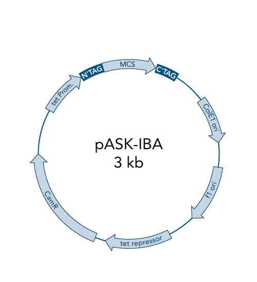
Features and benefits of the pASK-IBA vectors:
- High-level expression in E. coli
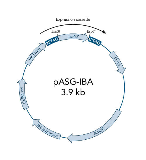
- Tightly regulated expression due to the tetracycline promoter
- Enhanced stability of cytotoxic genes
- Inexpensive induction with anhydrotetracycline
The pASK-IBA vectors are available with Strep-tag®II or 6xHis-tag, the OmpA secretion signal, special protease cleavage sites or Chloramphenicol resistance.
Principle and properties
pASK-IBA vectors work with the tightly regulated tetracycline (tet) promoter. Expression of the foreign gene is stringently repressed until induction with a low concentration of the chemical anhydrotetracycline. In contrast to the lac promoter, the tetA promoter/operator is tightly controlled and not functionally coupled to any cellular regulation mechanisms or genetic background. Unlike with the T7 promoter, special E. coli strains or extra plasmids are not required. The vectors do not mediate resistance against tetracycline.
The tet repressor is encoded on the pASK-IBA plasmids and is constitutively expressed from the b-lactamase or the chloramphenicol acetyl transferase promoter, respectively. This special arrangement guarantees a balanced stochiometry between repressor molecules and plasmid copy number. Expression of the foreign gene is stringently repressed until efficient chemical induction with a low concentration of anhydrotetracycline. In contrast to the lac promoter - which is leaky, susceptible to catabolite repression (cAMP-level, metabolic state), and influenced by chromosomally encoded repressor molecules - the tetA promoter/operator is tightly controlled and not functionally coupled to any cellular regulation mechanisms or genetic background.
As a consequence, special E. coli
strains or extra plasmids are not required and a broad range of culture
media and conditions can be used. For example, glucose minimal media
and even the XL1-Blue bacterial strain, which carries an episomal copy
of the tetracycline resistance gene, can be used for expression. The
pASK-IBA expression system is stable under many conditions, including fermentation, and is easy-to-handle.
Further
elements of the vectors are a tandem ribosome binding site (RBS) which
ensures efficient initiation of translation, the strong terminator of
the lipoprotein gene in order to prevent read-through, the intergenic
region of the bacteriophage f1 which provides a means for preparing
ssDNA and a b-lactamase or chloramphenicol acetyl transferase gene*. The vectors do not mediate resistance against tetracycline.
*Using cloning vectors with b-lactamase
resistance gene may be associated with some limitations since ampicillin
is degraded quite fast in bacterial culture medium. Therefore, we are
now offering our Strep-tag® II vectors pASK-IBA2C to pASK-IBA7C with chloramphenicol resistance instead of ampicillin resistance.
Reference:
Skerra, A. (1994). Use of the tetracycline promoter for the tightly regulated production of a murine antibody fragment in Escherichia coli. Gene, 151, 131-135.
The following E. coli strains have already been used successfully for Tet expression with our pASK-IBA vectors:
- JM83
- WK6
- B
- BL21
- MG1655
- W3110
- BL21(DE3)
- BLR(DE3)
- XL1-Blue
- BL21-CodonPlusTM-RIL
For secretion, we recommend JM83. For cytoplasmic expression E. coli
B strains are recommended, since they lack the lon protease and the
ompT outer membrane protease that can degrade proteins during
purification (Grodberg and Dunn, 1988, J.Bacteriol. 170, 1245).
Please note that we are not aware of an E. coli strain that is incompatible with the Tet expression system.
Anhydrotetracycline: Inducer for tetA Promoter
The E. coli expression cassette of the Strep-tag®/Strep-Tactin® over-expression system is under transcriptional control of the tetA promoter/operator and repressor. The promoter is induced by a low concentration of anhydrotetracycline (AHT) saving costs and minimizing the antibacterial influence of AHT. Degenkolb et al. (1991) have shown, that AHT binds 35-times tighter than tetracycline to the tet repressor.
Features and benefits of the pPSG-IBA vectors
- High-level expression by bacteriophage T7 promoter with pPSG-IBA vectors
- High-level transcription by T7 RNA polymerase in BL21 strains
- High-level expression of non-toxic proteins
- Induction by IPTG
- Universal cloning strategy with one restriction enzyme
- Suitable for in vitro transcription/translation
The pPSG-IBA vectors are also available with Twin-Strep-tag®, 6xHis-tag, GST-tag, Flag-tag or with two tags.
Principle and properties
The pPSG-IBA expression vectors are based on the T7 expression system. This system uses the T7 promoter and T7 RNA polymerase for high-level transcription of the gene of interest. Expression of the target genes is induced by providing a source of T7 RNA polymerase gene. The latter is under control of the lacUV5 promoter which can be induced by IPTG.
The advantage over the tet expression system is that this promoter is even stronger.
The Tet promoter is of medium strength which leads to high level expression of
certain proteins depending e.g. on their folding rate and stability –
characteristics which can hardly be predicted. Some proteins, however, can only
be expressed at high level if transcribed by a stronger promoter. In such cases
we recommend the use of the T7 promoter.
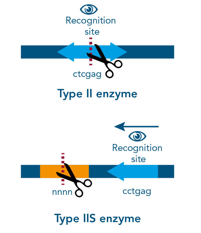
Type II enzymes are one of the 4 (I-IV) types of recognized endonucleases, which cut DNA at a particular recognition site. Type II enzymes cleave within or a short distance from their recognition sites, which comprise usually 4–8 nucleotides in length.
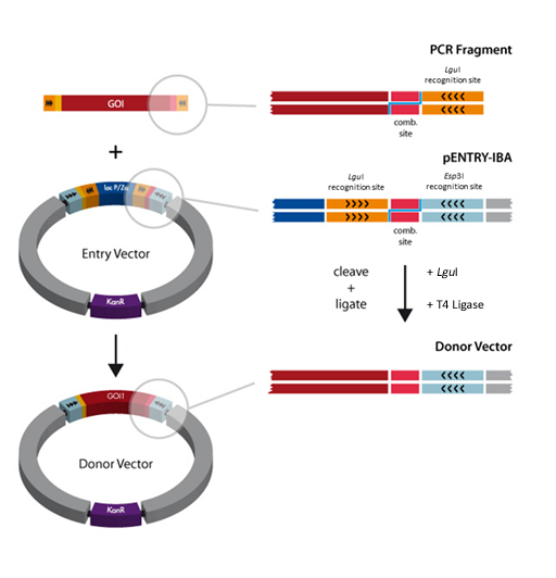
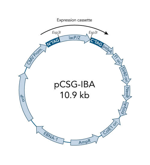
Among them, are the type IIS enzymes, like LguI and Esp3I.
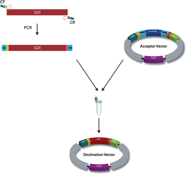
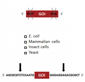
Type IIS restriction enzymes are dimeric enzymes that cleave DNA at a defined distance from their non-palindromic, asymmetric recognition site. This means that the target sequence can only be read in one direction. Thereby the digestion with only one single enzyme can generate two different independent sticky ends with 5’-overhangs allowing directional cloning. In addition, after digestion reaction the recognition sequence is removed completely and therefore the encoded amino acid sequence is not affected by remaining restriction enzyme sites.
The usage of typeIIS restriction enzymes provides important features for cloning:
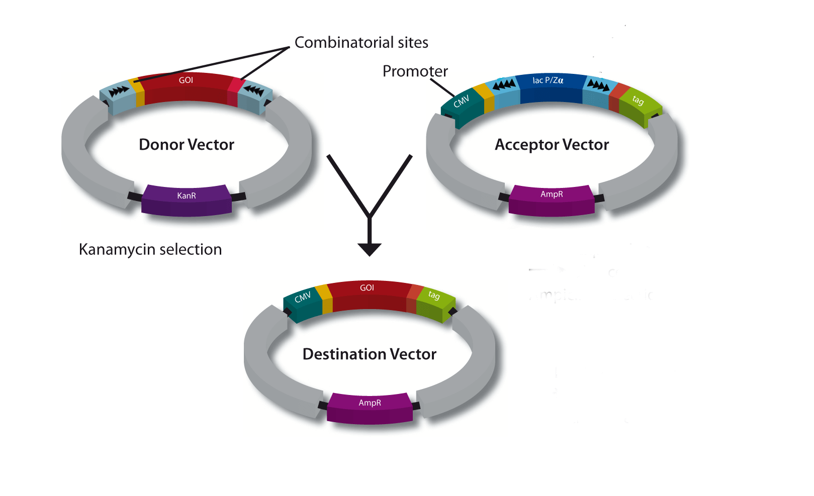
- It allows one tube cloning
- Expression of authentic proteins is possible (no additional amino acids)
- The cloning will be always in frame with the vector features
- Assembly of multiple fragments is possible
References
- Pingoud A, Jeltsch A (2001). Structure and function of type II restriction endonucleases. Nucleic Acids Res. 29 (18): 3705–27.